HsaEX0066152 @ hg38
Exon Skipping
Gene
ENSG00000041982 | TNC
Description
tenascin C [Source:HGNC Symbol;Acc:HGNC:5318]
Coordinates
chr9:115042219-115073866:-
Coord C1 exon
chr9:115073603-115073866
Coord A exon
chr9:115046410-115046682
Coord C2 exon
chr9:115042219-115042341
Length
273 bp
Sequences
Splice sites
3' ss Seq
ACCATTTTCTCTCCCTCTAGAAG
3' ss Score
11.02
5' ss Seq
CAGGTACTG
5' ss Score
9.04
Exon sequences
Seq C1 exon
AGTTGGACACGCCCAAGGACCTTCAGGTTTCTGAAACTGCAGAGACCAGCCTGACCCTGCTCTGGAAGACACCGTTGGCCAAATTTGACCGCTACCGCCTCAATTACAGTCTCCCCACAGGCCAGTGGGTGGGAGTGCAGCTTCCAAGAAACACCACTTCCTATGTCCTGAGAGGCCTGGAACCAGGACAGGAGTACAATGTCCTCCTGACAGCCGAGAAAGGCAGACACAAGAGCAAGCCCGCACGTGTGAAGGCATCCACTG
Seq A exon
AAGCCGAACCGGAAGTTGACAACCTTCTGGTTTCAGATGCCACCCCAGACGGTTTCCGTCTGTCCTGGACAGCTGATGAAGGGGTCTTCGACAATTTTGTTCTCAAAATCAGAGATACCAAAAAGCAGTCTGAGCCACTGGAAATAACCCTACTTGCCCCCGAACGTACCAGGGACATAACAGGTCTCAGAGAGGCTACTGAATACGAAATTGAACTCTATGGAATAAGCAAAGGAAGGCGATCCCAGACAGTCAGTGCTATAGCAACAACAG
Seq C2 exon
CCATGGGCTCCCCAAAGGAAGTCATTTTCTCAGACATCACTGAAAATTCGGCTACTGTCAGCTGGAGGGCACCCACAGCCCAAGTGGAGAGCTTCCGGATTACCTATGTGCCCATTACAGGAG
VastDB Features
Vast-tools module Information
Secondary ID
ENSG00000041982_MULTIEX1-8/8=C1-C2
Average complexity
ME(2-8[95=100])
Mappability confidence:
78%=78=100%
Protein Impact
Alternative protein isoforms (Ref)
No structure available
Features
Disorder rate (Iupred):
C1=0.301 A=0.115 C2=0.000
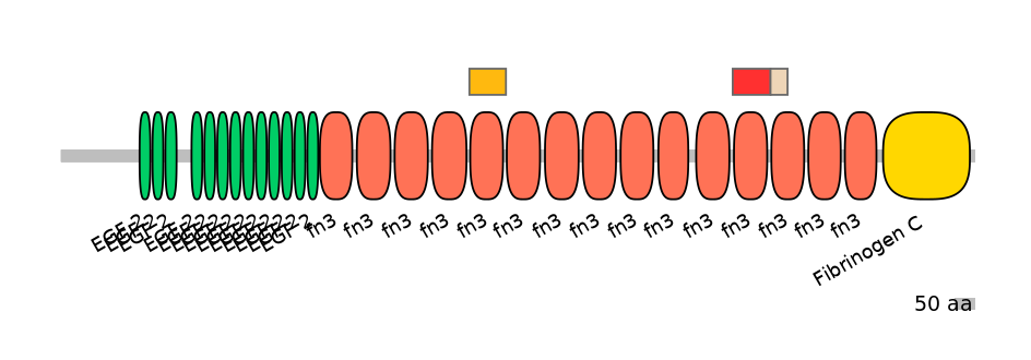
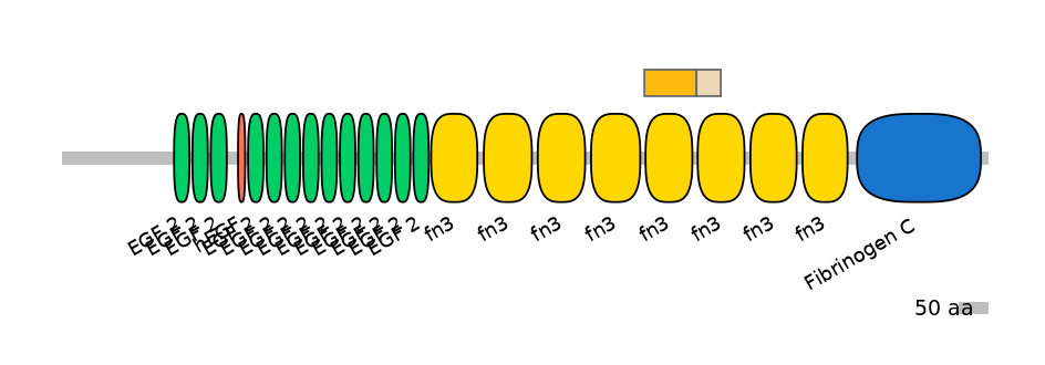
Domain overlap (PFAM):
C1:
PF0004116=fn3=WD(100=89.9)
A:
PF0004116=fn3=WD(100=88.0)
C2:
PF0004116=fn3=PU(48.8=92.9)


Associated events
Other assemblies
Conservation
Fruitfly
(dm6)
No conservation detected
Primers PCR
Suggestions for RT-PCR validation
F:
GACCGCTACCGCCTCAATTAC
R:
TGGGCACATAGGTAATCCGGA
Band lengths:
292-565
Functional annotations
There are 13 annotated functions for this event
PMID: 7499434
The small splice variant of tenascin-C (TNC) has eight fibronectin type III (FN3) domains (it lacks all alternatively splice exons, including: HsaEX0066148, HsaEX0066149, HsaEX0066151, and HsaEX0066152). The major large splice variant has three (in chicken) or seven (in human) additional FN3 domains (encoded by the alternative exons A1 (HsaEX0066148), A2, A3 (HsaEX0066149), A4, B,C (HsaEX0066151), D (HsaEX0066152)) inserted between domains five and six. Chiquet-Ehrismann et al. (Chiquet-Ehrismann, R., Matsuoka, Y., Hofer, U., Spring, J., Bernasconi, C., and Chiquet, M. (1991, Cell Regul. 2, 927-938) demonstrated that the small variant bound preferentially to fibronectin in enzyme-linked immunosorbent assay, and only the small variant was incorporated into the matrix by cultures of chicken fibroblasts. In this paper the authors have studied human tenascin-C, and confirmed that the small variant binds preferentially to purified fibronectin and to fibronectin-containing extracellular matrix. Thus this differential binding appears to be conserved across vertebrate species. Using bacterial expression proteins, they mapped the major binding site to the third FN3 domain of TNC. Consistent with this mapping, a monoclonal antibody against an epitope in this domain did not stain TNC segments bound to cell culture matrix fibrils. The enhanced binding of the small TN variant suggests the existence of another, weak binding site probably in FN3 domains 6-8, which is only positioned to bind fibronectin in the small splice variant. This binding of domains 6-8 may involve a third molecule present in matrix fibrils, as the enhanced binding of small TN was much more prominent to matrix fibrils than to purified fibronectin. Note, the function of individual exons were not examined.
PMID: 10367733
This study investigated the impact of cellular environment on the neurite outgrowth promoting properties of the alternatively spliced fibronectin type-III region (fnA-D) of tenascin-C. The effects of the A1-A4 region (consists of exon A1 (HsaEX0066148), A2, A3 (HsaEX0066149), and A4), B-D region (consists of exon B, exon C (HsaEX0066151), and exon D (HsaEX0066152) and the entire region A-D (all the mentioned exons) were examined. FnA-D promoted neurite outgrowth in vitro when bound to the surface of BHK cells or cerebral cortical astrocytes, but the absolute increase was greater on astrocytes. In addition, different neurite outgrowth promoting sites were revealed within fnA-D bound to the two cellular substrates. FnA-D also promoted neurite outgrowth as a soluble ligand; however, the actions of soluble fnA-D were not affected by cell type. It was hypothesized that different mechanisms of cellular binding can alter the growth promoting actions of bound fnA-D. The authors found that fnA-D utilizes two distinct sequences to bind to the BHK cell surface as opposed to the BHK extracellular matrix. In contrast, only one of these sequences is utilized to bind to the astrocyte matrix as opposed to the astrocyte surface. Furthermore, Scatchard analysis indicated two types of receptors for fnA-D on BHK cells and only one type on astrocytes. These results suggest that active sites for neurite outgrowth within fnA-D are differentially revealed depending on cell-specific fnA-D binding sites.
PMID: 10493745
Tenascin-C has been implicated in regulation of both neurite outgrowth and neurite guidance. A particular region of tenascin-C has powerful neurite outgrowth-promoting actions in vitro. This region consists of the alternatively spliced fibronectin type-III (FN-III) repeats A?D and is abbreviated fnA-D. The purpose of this study was to investigate whether fnA-D also provides neurite guidance cues and whether the same or different sequences mediate outgrowth and guidance. The examined exons were A1 (HsaEX0066148), A2, A3 (HsaEX0066149), A4, C (HsaEX0066151), and D (HsaEX0066152). The authors developed an assay to quantify neurite behavior at sharp substrate boundaries and found that neurites demonstrated a strong preference for fnA-D when given a choice at a poly-l-lysine?fnA-D interface, even when fnA-D was intermingled with otherwise repellant molecules. Furthermore, neurites preferred cells that overexpressed the largest but not the smallest tenascin-C splice variant when given a choice between control cells and cells transfected with tenascin-C. The permissive guidance cues of large tenascin-C expressed by cells were mapped to fnA-D. Using a combination of recombinant proteins corresponding to specific alternatively spliced FN-III domains and monoclonal antibodies against neurite outgrowth-promoting sites, the authors demonstrated that neurite outgrowth and guidance were facilitated by distinct sequences within fnA-D.
PMID: 11549732
The region of tenascin-C containing only alternately spliced fibronectin type-III repeat D (=HsaEX0066152), called fnD, when used by itself, dramatically increases neurite outgrowth in culture. Treatment of neuronal cells with alternatively spliced exon of tenascin-C promotes neurite growth. Study does not examine the function of any endogenous tenascin-C isoforms
PMID: 11714809
[Mapping: domain TnFnIII D; PMID:25482829]. Together, these 6-8 alternative exon array the encode minimal region of tenascin-C that can inhibit T cell activation. Recombinant fragments corresponding to defined regions of the molecule were tested for their ability to inhibit in vitro activation of human peripheral blood T cells induced by anti-CD3 mAbs in combination with fibronectin or IL-2. A recombinant protein encompassing the alternatively spliced fibronectin type III domains of tenascin-C (TnFnIII A1-3 and B-D) vigorously inhibited both early and late lymphocyte activation events including activation-induced TCR/CD8 down-modulation, cytokine production, and DNA synthesis. In agreement with this, full length recombinant tenascin-C containing the alternatively spliced region suppressed T cell activation, whereas tenascin-C lacking this region did not. Using a series of smaller fragments and deletion mutants issued from this region, the authors have identified the TnFnIII A1A2 domain as the minimal region suppressing T cell activation. Single TnFnIII A1 or A2 domains were no longer inhibitory, while maximal inhibition required the presence of the TnFnIII A3 domain.
PMID: 14715956
Neurite outgrowth by the alternatively spliced region of human tenascin-C is mediated by neuronal alpha7beta1 integrin.
PMID: 19405959
The stromal microenvironment has a profound influence on tumour cell behaviour. In tumours, the extracellular matrix (ECM) composition differs from normal tissue and allows novel interactions to influence tumour cell function. The ECM protein tenascin-C (TNC) is frequently up-regulated in breast cancer and the same authors have previously identified two novel isoforms - one containing exon 16 (TNC-16) (the exon is also called exon D (HsaEX0066152) and one containing exons 14 (exon B) plus 16 (TNC-14/16). The present study has analysed the functional significance of this altered TNC isoform profile in breast cancer. TNC-16 and TNC-14/16 splice variants were generated using PCR-ligation and over-expressed in breast cancer cells (MCF-7, T47D, MDA-MD-231, MDA-MB-468, GI101) and human fibroblasts. The effects of these variants on tumour cell invasion and proliferation were measured and compared with the effects of the large (TNC-L) (containing alternatively spliced exons A1 (HsaEX0066148), A2, A3 (HsaEX0066149), A4, B, C (HsaEX0066151), D) and fully spliced small (TNC-S) isoforms (skips all cassette exons). TNC-16 and TNC-14/16 significantly enhanced tumour cell proliferation (P < 0.05) and invasion, both directly (P < 0.01) and as a response to transfected fibroblast expression (P < 0.05) with this effect being dependent on tumour cell interaction with TNC, because TNC-blocking antibodies abrogated these responses. An analysis of 19 matrix metalloproteinases (MMPs) and tissue inhibitor of matrix metalloproteinases 1 to 4 (TIMP 1 to 4) revealed that TNC up-regulated expression of MMP-13 and TIMP-3 two to four fold relative to vector, and invasion was reduced in the presence of MMP inhibitor GM6001. However, this effect was not isoform-specific but was elicited equally by all TNC isoforms.
PMID: 19508743
Tenascin-C (Tnc) is an astrocytic multifunctional extracellular matrix (ECM) glycoprotein that potentially promotes or inhibits neurite outgrowth. To investigate its possible functions for retinal development, explants from embryonic day 18 (E18) rat retinas were cultivated on culture substrates composed of poly-d-lysine (PDL), or PDL additionally coated with Tnc or laminin (LN)-1, which significantly increased fiber length. When combined with LN, Tnc induced axon fasciculation that reduced the apparent number of outgrowing fibers. In order to circumscribe the stimulatory region, Tnc-derived fibronectin type III (TNfn) domains fused to the human Ig-Fc-fragment TNfnD6-Fc (Exon D (HsaEX0066152) and constitutive exon 6), TNfnBD-Fc (Exon B and Exon D), TNFnA1A2-Fc (Exon A1 (HsaEX0066148) and Exon A2) and TNfnA1D-Fc (Exon A1 and Exon D) were studied. The fusion proteins TNfnBD-Fc and to a lesser degree TNfnA1D-Fc were stimulatory when compared with the Ig-Fc-fragment protein without insert. In contrast, the combination TNfnA1A2-Fc reduced fiber outgrowth beneath the values obtained for the Ig-Fc domain, indicating potential inhibitory properties. The monoclonal J1/tn2 antibody (clone 578) that is specific for domain TNfnD blocked the stimulatory properties of the TNfn-Fc fusions. When postnatal day 7 retinal ganglion cells were used rather that explants, Tnc and Tnc-derived proteins proved permissive for neurite outgrowth.
PMID: 20678196
Tenascin-C (TNC) is a large extracellular matrix glycoprotein that shows prominent stromal expression in many solid tumours. The profile of isoforms expressed differs between cancers and normal breast, with the two additional domains AD1 and AD2 (encoded by HsaEX0066151) considered to be tumour associated. The aim of the present study was to investigate expression of AD1 and AD2 in normal, benign and malignant breast tissue to determine their relationship with tumour characteristics and to perform in vitro functional assays to investigate the role of AD1 in tumour cell invasion and growth. Expression of AD1 and AD2 was related to hypoxanthine phosphoribosyltransferase 1 as a housekeeping gene in breast tissue using quantitative RT-PCR, and the results were related to clinicopathological features of the tumours. Constructs overexpressing an AD1-containing isoform (TNC-14/AD1/16) (contains the cassette exons B, AD1, and D (HsaEX0066152) were transiently transfected into breast carcinoma cell lines (MCF-7, T-47 D, ZR-75-1, MDA-MB-231 and GI-101) to assess the effect in vitro on invasion and growth. Statistical analysis was performed using a nonparametric Mann-Whitney test for comparison of clinicopathological features with levels of TNC expression and using Jonckheere-Terpstra trend analysis for association of expression with tumour grade. Quantitative RT-PCR detected AD1 and AD2 mRNA expression in 34.9% and 23.1% of 134 invasive breast carcinomas, respectively. AD1 mRNA was localised by in situ hybridisation to tumour epithelial cells, and more predominantly to myoepithelium around associated normal breast ducts. Although not tumour specific, AD1 and AD2 expression was significantly more frequent in carcinomas in younger women (age ?40 years; P < 0.001) and AD1 expression was also associated with oestrogen receptor-negative and grade 3 tumours (P < 0.05). AD1 was found to be incorporated into a tumour-specific isoform, not detected in normal tissues. Overexpression of the TNC-14/AD1/16 isoform significantly enhanced tumour cell invasion (P < 0.01) and growth (P < 0.01) over base levels.
PMID: 32599145
The pro-inflammatory activity of an endogenous innate immune trigger is controlled by inclusion or exclusion of a novel immunomodulatory site located within domains AD2AD1 (both exons), identifying this as a mechanism that prevents unnecessary inflammation in healthy tissues but enables rapid immune cell mobilization and activation upon tissue damage, and defining how this goes awry in autoimmune disease.
PMID: 1725601
Three consecutive exons encoding one FN3 domain each. Modulation of binding to Fibronectin.
PMID: 12151539
Tenascin-C is a multimodular glycoprotein that possesses neurite outgrowth-stimulating properties, and one functional site has been localized to the alternatively spliced fibronectin type III domain D (encoded by HsaEX0066152). To identify the neuronal receptor that mediates this effect, neighboring pairs of fibronectin type III domains: exon A1 (HsaEX0066148), exon A2, exon B, exon C (HsaEX0066151), exon D (HsaEX0066152), were expressed as hybrid proteins fused to the Fc fragment of human immunoglobulin. These IgFc fusions were tested for neurite outgrowth-promoting properties on embryonic day 18 rat hippocampal neurons, and both the combinations BD and D6 were shown to promote the elongation of the longest process, the prospective axon. Antibodies to the cell adhesion molecule F3/contactin of the Ig superfamily blocked the BD- but not the D6-dependent effect. Biochemical studies using F3/contactin?IgFc chimeric proteins confirmed that the adhesion molecule selectively reacts with the combination BD but not with other pairs of fibronectin type III repeats of tenascin-C. The alternatively spliced BD cassettes are prominently expressed in the developing hippocampus, as shown by reverse transcription PCR, and colocalize with F3 expression during perinatal periods when axon growth and the establishment of hippocampal connections take place. Note, the exons are tested in combination with one another, e.g. B and D are tested together. The effect of individual exons are not tested in isolation.
PMID: 19394429
Tenascin-C (Tnc) is transiently expressed during neural development. Within its alternatively spliced fibronectin type III (TNfn) -motifs the TNfnD domain (encoded by HsaEX0066152) is crucial for a neurite outgrowth-promoting region that is recognized by the GPI-linked adhesion molecule of the Ig-superfamily contactin. In order to understand the downstream signaling mechanisms, embryonic day E18 rat hippocampal neurons were cultivated on TNfnBD-containing and control substrates in the presence of various inhibitors. As predicted, axon outgrowth promotion could be suppressed by antibodies to the TNfnD domain, to contactin, or to the beta1-integrin subunit. The chelators BAPTA/AM or EGTA as well as blockade of membrane-based calcium channels or of the release of calcium from intracellular stores reduced axon growth to control levels. The inhibition of phospholipase C and its downstream targets protein kinase C or calmodulin kinase likewise blocked outgrowth promotion.
GENOMIC CONTEXT[edit]
INCLUSION PATTERN[edit]
SPECIAL DATASETS
- The Cancer Genome Atlas (TCGA)
- Genotype-Tissue Expression Project (GTEx)
- Autistic and control brains
- Pre-implantation embryo development